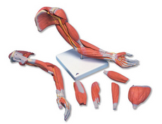
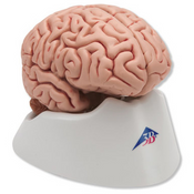
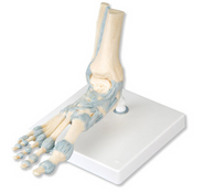
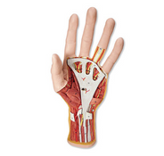
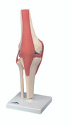
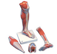
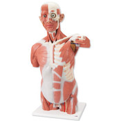
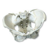
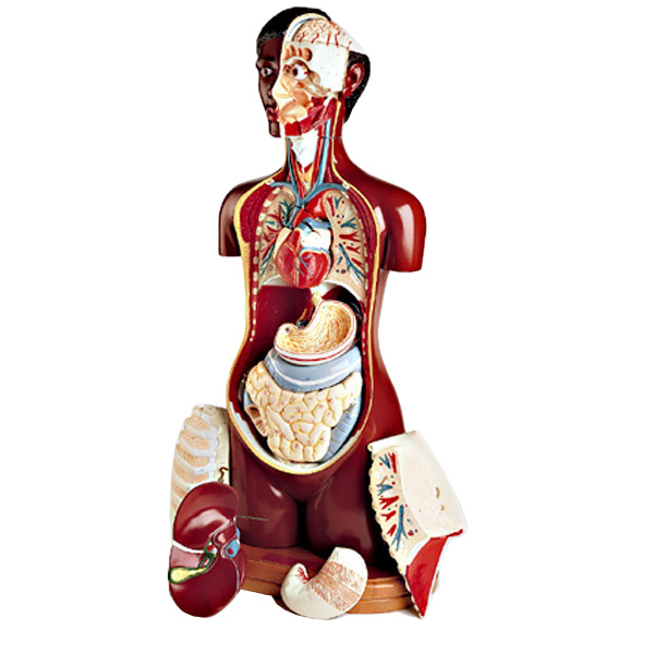
3-B Scientific produces a variety of anatomical models from full body skeletons to individual joints and specific body parts. These meticulously designed, durable made and manufactured models are designed to provide the educator and the student with the ability to demonstrate and visualize the human anatomy in the unique 3-dimensional perspective of being handled, turned around, disassembled, and reassembled for an enhanced learning experience.
Models: arm, leg, brain, ear, eye, foot, hand, heart, larynx, joints, spine, skull, pelvis, muscles and bones.


